In the early foetal period, the cerebrum covers about half of the midbrain. The separation of the dural limiting layer in the parieto-occipital region extends from the posterior cerebrum to the cranial cerebellum. The lateral folds of the tentorium cerebelli were distributed between its apex, which coincides with the falx cerebri, and its base plane, which is located between the midbrain and the rostral hindbrain.
The differences in the growth directions of the tentorium cerebelli components gradually diminish as the cerebrum covers the midbrain. A rotation of the tentorium cerebelli in its middle section was demonstrated, corresponding to its growth ceasing in the mid-fetal period.
The brainstem and cerebellum expanded downwards through differential growth, while the cerebrum covered them upwards. The morphology of the tentorium cerebelli curved to fit the cerebellar and cerebral surfaces.
A study by Matsunari et al. 2022 suggests that the morphology of the tentorium cerebelli is influenced by factors that differ particularly in the early and mid-fetal periods.
The present data from Matsunari et al. 2022 show a much more comprehensive picture of tentorium cerebelli formation as a function of fetal developmental stage than has previously been presented.
The following is the timetable of morphogenesis of the embryonic and foetal brain and tentorium cerebelli (Matsunari et al. 2023).

Abbreviations:
Cb: Cerebellum;
CRL: Crown-rump length,
CS: Carnegie Stadium;
Cx: Cerebrum;
DS: Dorsum sellae;
FC: Falx cerebri;
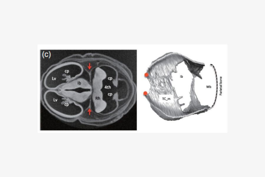
Ib: Diencephalon;
iv_f: first
Involvement in the parieto-occipital region;
Mb, midbrain;
Rh, rostral hindbrain;
SNL,
upper neck line;
TC, Tentorium cerebelli;
pCx: posterior end of the cerebrum;
tTC: most cranial part of the cerebellum;
bTC: Point at which the posterolateral part of the TC becomes wide (here the double-layered dura mater is divided into its cranial and caudal components);
cCb: the most cranial part of the cerebellum
To [1] The angle pCx_tTC had a negative value.
Re [2] The iv_f and lateral TC appear separately and extend in different directions.
To [3] These phenomena were correlated.
Re [4] Two depressions and one invagination of the TC; the orientations of the iv_f and lateral TC appear different.
Re [5] The angle pCx_cCb and bTC_cCb had a negative value. (CRL >140 mm).
Matsunari C, Kanahashi T, Otani H, Imai H, Yamada S, Okada T, Takakuwa T. Tentorium cerebelli formation during human embryonic and early fetal development. Anat Rec (Hoboken). 2023 Mar;306(3):515-526.
https://pubmed.ncbi.nlm.nih.gov/36326822/