Aus Liem T. Van den Heede P. Foundations of Morphodynamics in Osteopathy: An Integrative Approach to Cranium, Nervous System, and Emotions, 2017; 1. Aufl. Handspring Publishing Limited, Pencaitland.
The contemporary mathematician L. Edwards studied the development of shape mainly in association with vortex patterns, extending his investigations into the development of embryological shape.
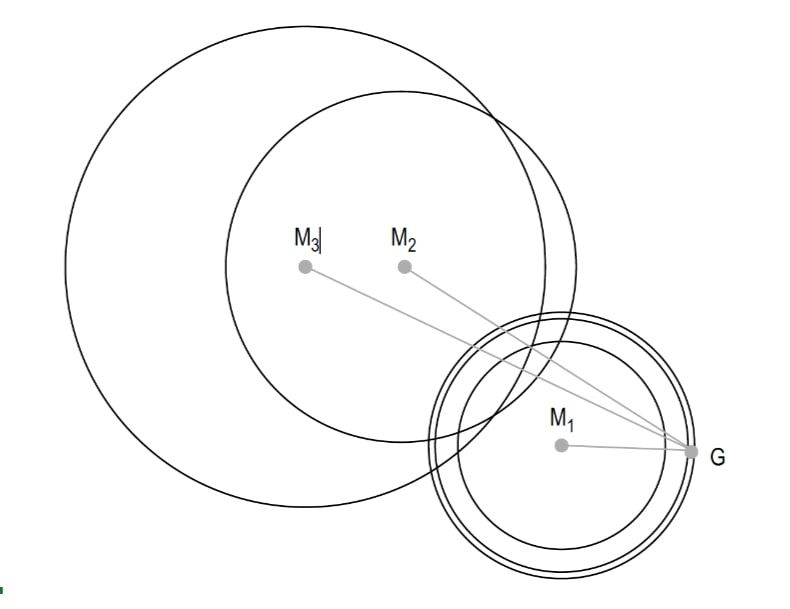
He sees the emergence of embryological shape as the perfect creation of a spiritual universe, which assumes shape according to the laws of mathematics. He addressed among other issues the question as to what form the geometrical calculations might produce if the axis of the airy vortex lay asymmetrically in relation to the axis of the bud transformation of a plant bud. For this calculation he selected six airy vortices from a common field. Their common axis was placed parallel to the axis of the bud transformation, but shifted to one side. Taking the graphic representation of these calculations, and evaluating various cross-sections, produced a clear resemblance to the invagination process that takes place during the development of the mesoderm in the third week of gestation. Even when the parameters were varied, for example by moving the vortex or the orientation of the axes to each other, the result still resembled an embryonic form. These forms, viewed in sequence, are reminiscent of various stages of embryonic development. By using a simple movement of a vortex, transformed by “egg transformation” (a certain set of mathematical calculations), it became possible, for example, to describe the general shape of a developing embryo. A certain positioning of the vortex (and of the resulting transformations) produced a remarkable similarity to the general form of a developing embryo. If these calculation models are applied to the uterus – which, like the egg and the flower bud, exhibits a good path curve form in the first weeks of pregnancy – a clear relationship or similarity is found between the biological reality (the shape of the actual stage of development of the embryo) and the geometric cross-sections obtained from certain calculation processes (Fig. 1). Edwards postulates that the development of seeds and avian embryos can be traced to the shape of the bud and the egg (in the sense of an operative, active shaping force). Shape could then be demonstrated as an active force for (initial or subsequent) development by the general sequence of formative gestures in embryonic development, by means of these geometric transformation processes. In line with this thinking, Edwards tries to represent neural tube formation in geometric terms. A constant increase in the width of the vortex, as in graphic representations of neural tube formation, produces growth of the two sides toward each other until eventually they meet and fuse (Fig. 2). The widest vortex produces a shape that achieves the closing of the tube. All that is needed to produce this remarkable transformation geometrically is to have an airy vortex (transformed by uterus transformation, i.e., the increased curvature of the uterus at the beginning of pregnancy) increase its radius. Another possible way of achieving this result (with minor deviations) would be to retain the same radius and move the vortex, from above to below, from below to above, or inward from both sides. In order to convert the process of neural tube closure (beginning at the middle and proceeding cranially and caudally) into geometric terms, we need to think of two vortices approaching each other from opposite directions: from above to below and from below to above, or inward from right and left . The process of neural tube closure according to Edwards could be interpreted metaphorically as follows: the first vortex from the past, the second from the future; or, the first vortex communicating the unconscious element of our being and the second, the conscious.


Aus Liem T. Van den Heede P. Foundations of Morphodynamics in Osteopathy: An Integrative Approach to Cranium, Nervous System, and Emotions, 2017; 1. Aufl. Handspring Publishing Limited, Pencaitland.


